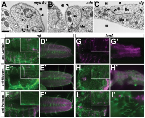
Figure 4. Exploring molecular mechanisms of Laminin A-dependent BM attachment.
Images of neuromuscular bouton profiles (A–C) in late stage 17 embryos carrying the following mutant allele combinations: mysXG43; ßInt-v1 in homozygosis (A), Sdc97/Sdc23 (B), Dg043/Dg086 (C); no changes in adhesion indices were detected (statistical validation in Fig. 2E); white arrows indicate pseudo-cell contacts separated by BMs, all other symbols as explained in Fig. 1. D–E′) Tissues of late stage 17 wildtype (left) or lanA9.32 mutant embryos (right): D–I) show flat-dissected whole body preparations (insets show the ventro-longitudinal muscles VL1-4) [109]; D′–H′) show isolated CNSs; I′ shows a close up of a flat dissected embryo; preparations are immuno-stained against Laminin, Nidogen or Perlecan (in green; as indicated on the left) in combination with anti-HRP labelling neuronal tissues (magenta). Perlecan and Nidogen are still present within fragmented BMs of lanA9.32-mutant embryos. Scale bar in A represents 600 nm in A, 200 nm in B and C, 80 µm in D–I (insets 2.5 fold enhanced), and 30 µm D–I′.

