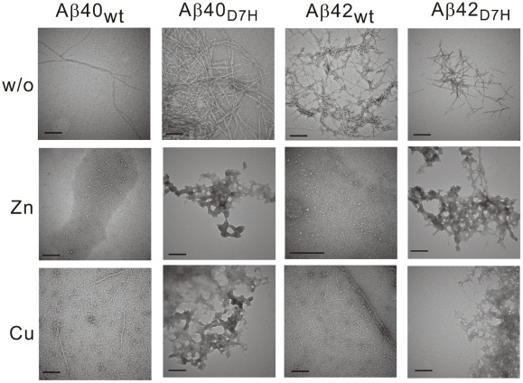
Figure 4. Aβ morphology in the presence or absence of metal ions was revealed by TEM.
Lyophilized Aβ was prepared in HFIP-DMSO. After 264–312 h of incubation in either the presence or absence of Zn2+ or Cu2+, the Aβ samples were stained by 2% uranyl acetate and monitored by TEM. In the presence of ions, the AβD7H peptides were predominantly amorphous morphology but not protofibrils as Aβwt. Scale bar: 200 nm.

