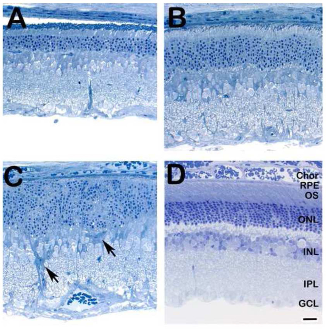
Figure 3.
Histological analysis of retinas from an adult rat one week after a single intravitreal injection with (A–C) 7-kChol (0.25 µmol, in 5 µl DMSO) or (D) DMSO alone (5 µl; vehicle control). Note the normal histological appearance of the retina from the vehicle control eye, compared to the marked retinal degeneration observed in the oxysterol-treated eye. Arrowheads (panel C) indicate gliotic elements in the degenerating retina. Spurr’s resin embedment, 1-µm thick sections, Toluidine blue stain. Abbreviations: Chor, choroid; RPE, retinal pigment epithelium; OS, photoreceptor outer segment layer; ONL, outer nuclear layer; INL, inner nuclear layer; IPL, inner plexiform layer; GCL, ganglion cell layer. Scale bar (all panels), 25 µm.

