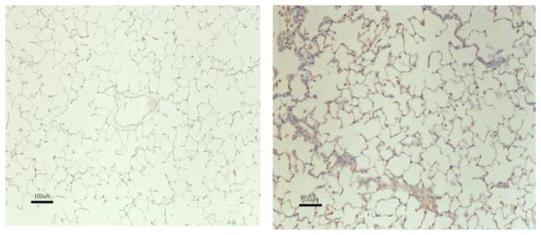
Figure 6. SM stimulates the production of TGF-β in the lung.
F344 rats were exposed to vehicle or SM (150 mg/m3) as described in “Materials and Methods.” At day 28 days post exposure, rats were sacrificed to collect lung tissues; IHC staining on lung sections was performed for TGF-β. Tissue sections treated with vehicle (left panel) and SM (right panel) were evaluated for TGF-β expression. Brown staining represents TGF-β reactivity. The figure is representative of 6 animals in each group.

