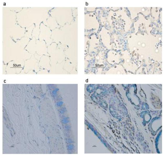
Figure 7. SM induces accumulation of IL-17+ cells in the lung.
F344 rats and cynomolgus macaques were exposed to vehicle or SM (150 mg/m3) as described in “Materials and Methods.” Lung sections were processed for intracellular IHC staining to visualize IL-17+ cells. The sections represent rat control (a), rat SM (b), monkey control (c), and monkey SM (d). Brown staining represents IL-17+ cells (right panels); vehicle control sections show minimal staining (left panels).

