
Fig. 5.
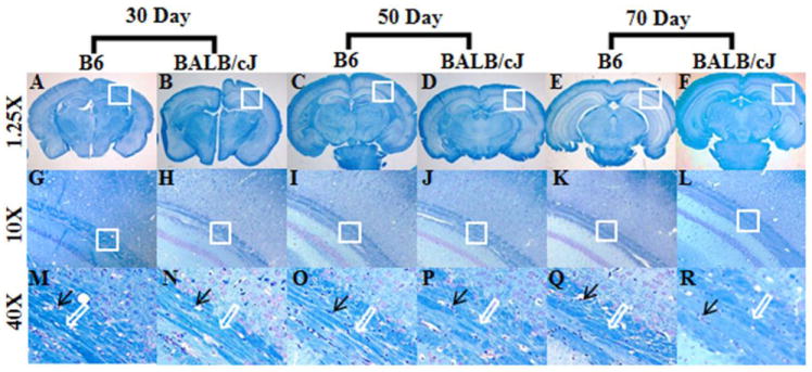
Representative luxol-fast blue (LFB) stained sections from the external capsule region in B6 and BALB/cJ brain samples. Figure A-F represents the brain sections at 1.25x resolution. Figure G-L showing images from the rectangular box from external capsule at a magnification of the 10X and figure M-R demonstrates further magnification at 40X from the rectangular box shown in G-L. Black arrow demonstrating the vacuolated nucleoli from the axonal fibers, while the white arrow indicates to the relative compactness of the white matter fibers.