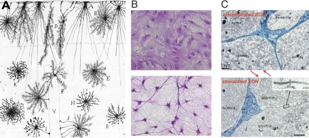
Figure 4. Morphological plasticity of astrocytes.

(A) Golgi-stained astrocytes from a 2 month-old human infant in cortical layers I-III. A, B, C, and D are cells in layer I, whereas E,F,G and H are astrocytes in layer II and II. I and J are cells with endfeet contacting blood vessels. V, blood vessel. (Ramón y Cajal, 1913). (B) Cultured rat astrocytes before (top panel) and after addition of dBcAMP (1 μM, lower panel), cresyl violet. Courtesy of Drs. Fujita and Abe, Hoshi University. (C) Electron microscopy of the rat SON during parturition (top panel) and lactation (lower panel). Astrocytes withdraw their processes in lactating animals. From (Theodosis et al. 2008).
