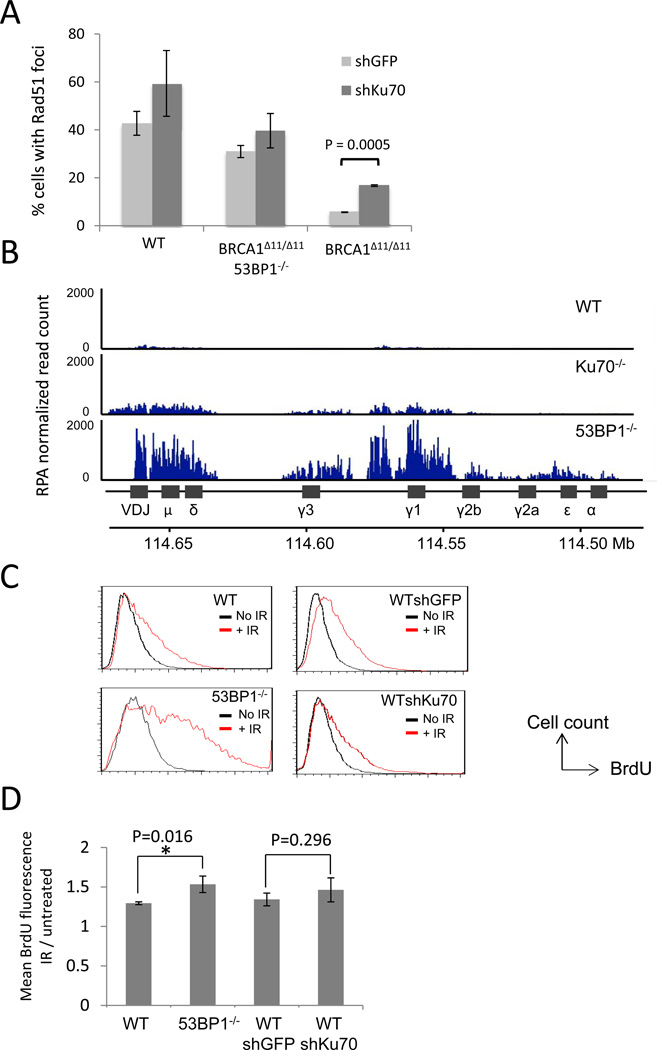
Figure 3. Effect of 53BP1 and Ku on Rad51 foci and DSB resection.
(A) Quantification of Rad51 immunofluorescence in mouse embryonic fibroblasts of the indicated genotypes. Cell expressing either control shRNA against GFP or shRNA against Ku70 were irradiated (5Gy, 4hrs recovery) and stained with anti-Rad51 antibody. The average percentages of cells (+/− standard deviation) with more than 5 nuclear foci from three experiments are shown. (B) Anti-RPA ChIP-Seq in B cells. B cells were stimulated to undergo class switch recombination in vitro. Chromatin from B cells was harvested 48 hrs post-stimulation and used for RPA ChIP. RPA read count (normalized by the total library size per million) is shown at the IgH locus. (C) Non-denaturing anti-BrdU immunofluorescence in MEFs treated with ionizing radiation (30Gy, 2 hrs recovery), measured by flow cytometry. Resection is measured by detection of exposed BrdU (x-axis). (D) Quantification of mean BrdU fluorescence intensity of the irradiated population shown in (C), normalized to the untreated population. Average +/− standard deviation from 5 experiments. See also Figure S4.

