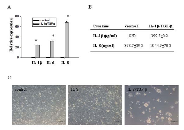
Figure 7.
Role of IL-1β, IL-6 and IL-8 in IL-1β/TGF-β-induced LN-229 neurosphere formation. (A) Relative gene expressions of IL-1β, IL-6, and IL-8 were determined by qRT-PCR. β-actin was used as an internal normalization control. Error bars represent SEM. *P < 0.05. (B) Secretion of IL-1β and IL-8 from control LN-229 cells and IL-1β/TGF-β-induced neurosphere cells. The same number of LN-229 cells was cultured in SFM with or without IL-1β/TGF-β for six days, and then culture media was removed. Cells were washed with PBS, and cultured in fresh SFM without IL-1β/TGF-β for 24 hours. The media were collected, followed by ELISA analysis. N/D: not detected. (C) Combination of IL-8 and TGF-β induce formation of neurospheres. LN-229 cells were cultured in SFM in the presence or absence of IL-8 or IL-8/TGF-β for seven days. Scale bar = 200 μm.

