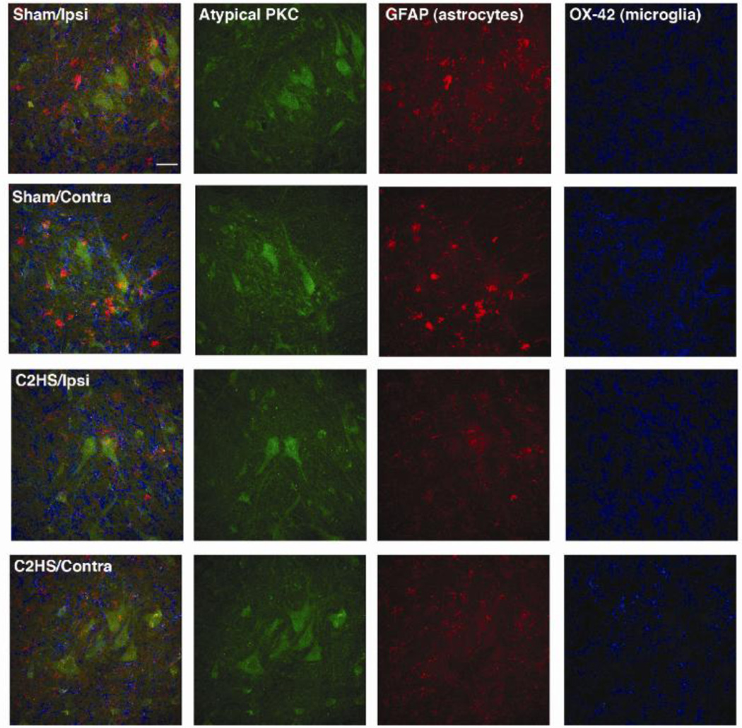
Figure 5.
Atypical PKC is expressed in presumptive phrenic motor neurons in sham operated and C2 hemisected rats. All images are from the ventrolateral region of the ventral horn at C4 28 days post-surgery. The left column of panels are the combined images of aPKC (green), GFAP (astrocytes, red) and OX-42 (microglia, blue). Scale bar = 40µm.

