
Figure 2.
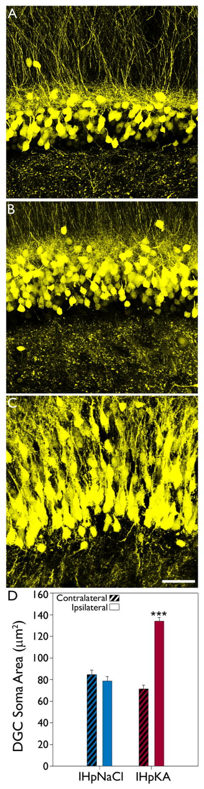
Intrahippocampal injection of kainic acid induces ipsilateral granule cell somatic hypertrophy. Representative confocal maximum projections of YFP-expressing granule cells located in the upper blade of dentate gyrus from the ipsilateral hemisphere of an IHpNaCl mouse (A), contralateral hemisphere of an IHpKA mouse (B) and ipsilateral hemisphere of an IHpKA mouse (C). Note the hypertrophied granule cell somata within the ipsilateral hemisphere of the IHpKA mouse. Scale bar = 40 μm. D: Mean area (± SEM) of YFP-expressing granule cell somata by treatment group and hemisphere. ***P<0.001 vs. all other groups.