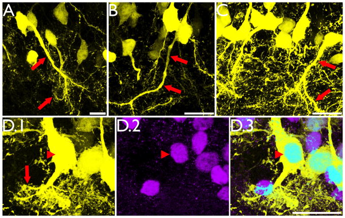
Figure 5.
Intrahippocampal injection of kainic acid results in the formation of hilar basal dendrites with structures similar to those observed in other models of epilepsy (A–C, red arrows) and novel “tufted” hilar basal dendrites (D.1, red arrow) on YFP-expressing granule cells. To confirm the granule cell identity of the cell shown in D.1, the section was co-labeled with the granule cell specific marker Prox1 (D.2). Colocalization of YFP and Prox1 is shown in D.3 (arrowhead). Images are confocal maximum projections. Scale bars = 20 μm.

