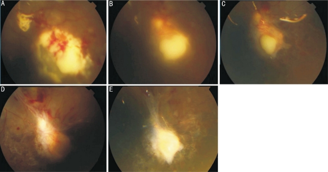
Figure 3. Fundus appearance-post vitrectomy.
A: 3rd day, the remaining lesion of inferior retina enlarged a little, multiple bleedings were seen across the retina, but satellite lesions were stable; B: 12th day, the yellowish-white lesion became localized; C: 19th day, the lesion with distinct borders became smaller, satellite lesions showed gradual recession; D: 28th day, the greyish-white lesion in inferior retina resolved and was not raised any more. Infratemporal retina showed strand of scarring with fibrosis from vascular arcades to focal zone. Greyish-yellow macular edema and scattered hemorrhagic foci were still present; E: 103th day, hemorrhagic foci resolved and a scar with well-demarcated borders remained

