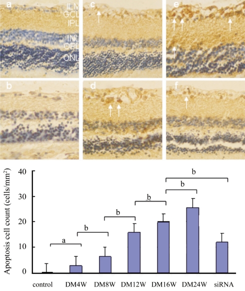
Figure 2. Tunnel positive cells (arrows) appeared in diabetic retina.
a: normal retina; b: diabetic 4 weeks; c: diabetic 8 weeks; d: diabetic 16 weeks; e: diabetic 24 weeks; f: diabetic 16 weeks interfered by CTGFsiRNA. The internal limiting membrane (ILM), the ganglion cell layer (GCL), the inner plexiform layer (IPL), the outer plexiform layer (OPL), and the outer nuclear layer (ONL).The difference is significant (aP <0.05, bP <0.01)

