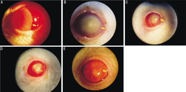
Figure 1. Clinical progression of F. solani-induced keratomycosis after inoculation with 108CFU/mL spore suspension of F. solani.
A: slitlamp photos showing corneal edema and infiltration after 1 day; B: yellow corneal ulcer with compact texture after 3 days; C: smaller corneal ulcer with epithelial recovery on the wound edge after 6 days; D: opaque cornea with more epithelial recovery after 10 days; E: complete corneal recovery after 14 days of the infection. Representative physical signs are shown as arrows pointed. The mock-inoculated corneas were normal after 3 days of infection

