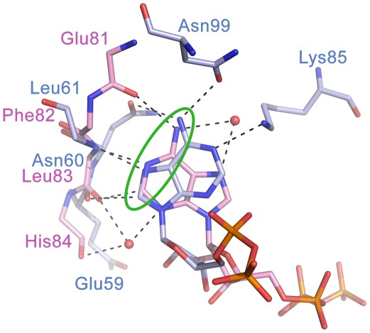
Figure 3. Superposition of adenine binding sites of human CDK2 and AaIspE.
ATP adopts the anti conformation in hCDK2 (PDB code 2cch, carbon atoms coloured pink) while the ATP analogue AMP-PNP adopts the syn conformation in AaIspE (PDB code 2v8p, carbon atoms coloured light blue). Further, in hCDK2 adenine forms hydrogen bonds with the backbone amide groups of the hinge region while in AaIspE the backbone and side chain atoms of surrounding amino acids are involved in hydrogen bonding-interactions. In both enzymes, a donor-acceptor-donor motive (green circle) is important for molecular recognition.

