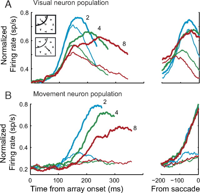
Figure 3.
Frontal eye field physiology during visual search. A, B, Normalized population firing rates for FEF visual (A) and movement (B) neurons during visual search with set size 2 (blue), 4 (green), and 8 (red). Spike density functions were generated from trials in which the target (thick) or a distractor (thin) was inside the RF of the neuron. Spike density functions are aligned on the stimulus array presentation (left) and saccade (right). Stimulus-aligned spike density functions end at the median RT.

