Fig. 7.
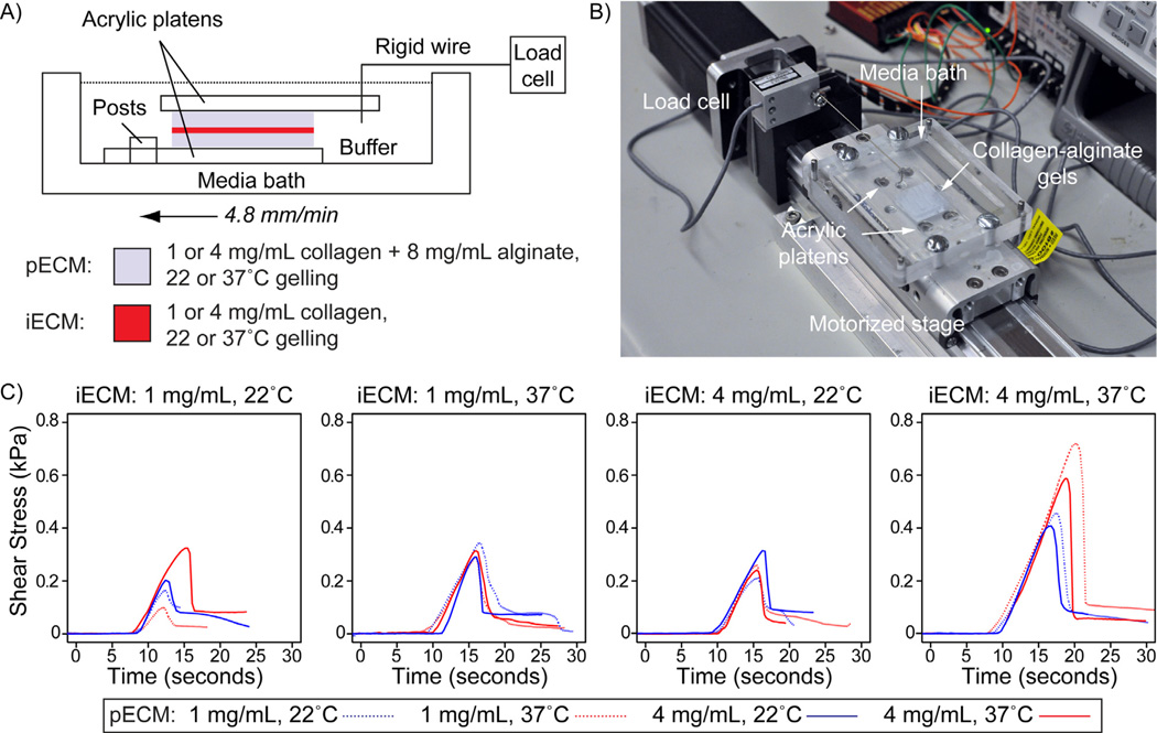
Characterization of interface adhesion strength by single lap shear adhesion testing. (A) Schematic of mechanical testing apparatus. Two collagen-alginate gels are glued to acrylic platens using cyanoacrylate, and then glued to each other with collagen. The gels on platens are then transferred to the media bath. Posts in the bath constrain the lower platen. A rigid steel wire connected to the load cell is inserted into a triangular cut in the top platen. A microcontroller controlled motorized stage is used to translate the specimen at 4.8 mm/min relative to the load cell. (B) Image of testing platform. (C) Representative shear adhesion test curves for each assembly condition. Each plot shows representative curves for each pECM assembly condition for a given iECM assembly condition.

