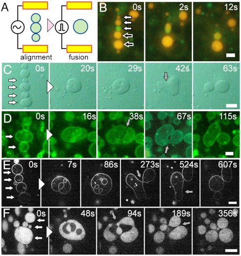
Fig. 1.
Electrofusion and budding transformation of GUVs. (A) Schematic representation of the electrofusion experimental setup. (B) Sequential epifluorescence images of the electrofusion of GUVs containing GFP (green) and R-PE (orange) without polymer. White and black-filled arrows at time zero indicate vesicles to fuse together. (C–F) Sequential images of budding transformations of vesicles containing 3 mM PEG 6000. White-filled arrows at time zero indicate vesicles to fuse together. Gray-filled arrows indicate the neck formation before budding. (C) Bright-field images. (D) Epifluorescence images of the membrane marked with fluorescence lipids. (E) Confocal fluorescence images of the membrane marked with fluorescence lipids. (F) Confocal fluorescence images of the vesicles encapsulating FITC-BSA. (Scale bars: 10 μm.)

