Fig. 2.
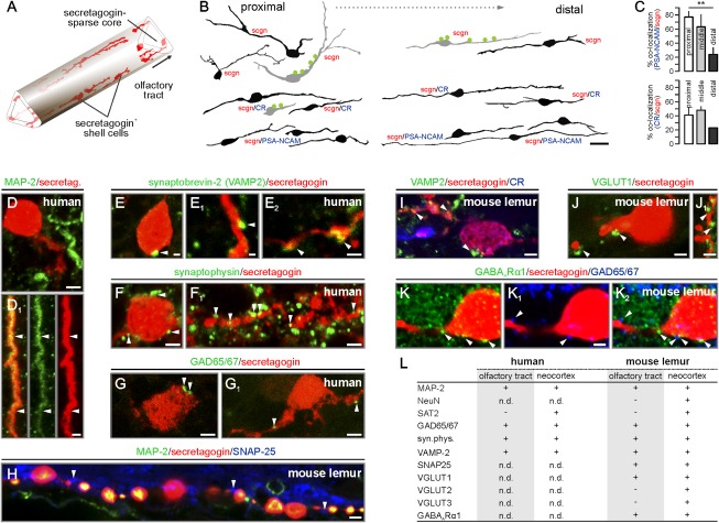
Secretagogin+ neurons of the olfactory tract synaptically integrate into the olfactory circuitry. (A) Secretagogin+ bipolar shell cells populate the margins of the human olfactory tract. (B) Secretagogin+ cells are predominantly bipolar, except a subset of multipolar calretinin (CR)−/PSA-NCAM− shell cells at locations proximal to the trigone. Somatodendritic afferents are in green. (C) The percentage of secretagogin+/CR+ and secretagogin+/PSA-NCAM+ neurons declines toward the distal olfactory tract. **P < 0.01. (D and D1) Dendritic (arrowheads), but not somatic MAP-2, immunoreactivity was seen in secretagogin+ neurons. (E–H) Synaptobrevin+ (E–E2), synaptophysin+ (F and F1), or GAD65/67+ (G and G1) profiles (arrowheads) on secretagogin+ somata and dendrites. (H–K) Similar to the human olfactory tract, SNAP25+ (H), synaptobrevin-2+ (I), VGLUT1+ (J and J1), or GAD65/67+ (K–K2) boutons contact secretagogin+ somata or processes in the mouse lemur olfactory tract. Note that postsynaptic GABAARα1 subunits (31) appose GAD65/67+ afferents (K2). (L) Markers of cellular identity and afferent synapses on secretagogin+ neurons (n.d., nondetectable due to e.g., epitope mismatch). [Scale bars: 10 μm (B), 3 μm (D, E2, F, G, and G1), 2 μm (D1, F1, H, I, I1, J, K, K1, and K2), and 1 μm (E and E1).]

