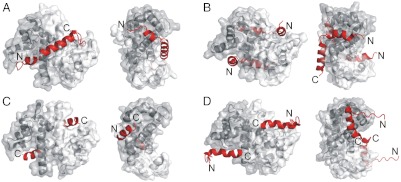
Fig. 6.
Comparison of four peptide-bound S100 proteins. The structure of (A) S100A4-NMIIA (3ZWH), (B) S100A6-SIP (2JTT) (25), (C) S100A10-ANXA2 (1BT6) (21), and (D) S100B-p53 (1DT7) (22) complexes are shown in two orientations. The left figures illustrate the two canonical hydrophobic pockets, while in the right ones the dimers are rotated to show the groove (the waist) formed by helices 3-3′ and 4-4′ that is occupied in the S100A4 complex by the central helical part of the myosin binding peptide. Peptide ligands are shown in red.

