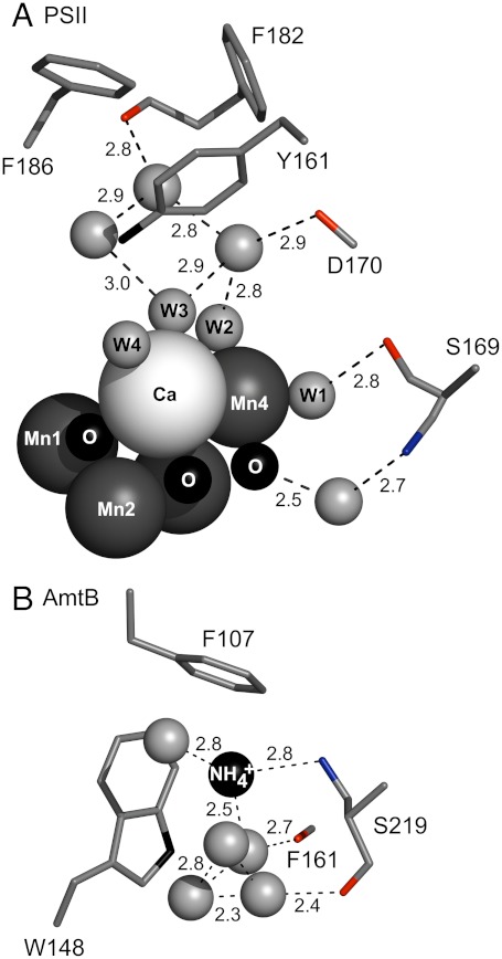
Fig. 8.
Similarities between the OEC and the ammonia binding site in the ammonia transport protein, AmtB. Fig. 8A was generated from the 1.9 Å structure of PSII (3). Fig. 8B was generated from the 1.35 Å structure of AmtB (15). Dashed lines show distances between atoms (Å). Amino acid residues are shown as sticks. Peptide carbonyl oxygen atoms are in red. The hydroxyl group of serine is in blue. Assigned water molecules in the OEC hydrogen-bonding network are denoted as W1-4 in (A). All other water molecules are shown as unlabeled gray spheres.

