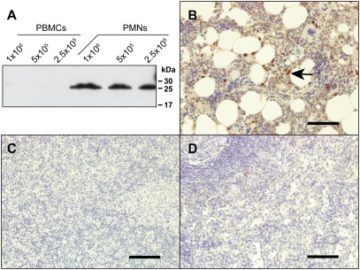
Fig. 3.
NSP4 is present in granulocytes. (A) Total cell lysates of peripheral blood mononuclear cells (PBMCs) and PMNs, which are almost exclusively neutrophil granulocytes, were analyzed by Western blotting using anti-NSP4 mAbs and secondary anti-rat HRP-labeled Ab. Natural NSP4 of PMNs runs lower than recombinant S-tag-NSP4 (Fig. 2B) because it is N-terminally processed and has shorter carbohydrate chains. (B–D) Staining of different human tissue samples using anti-NSP4 mAbs and the Ultravision LP staining kit. NSP4 was detected in neutrophil granulocytes and precursors in bone marrow tissue (arrow, B). NSP4 was not detected in lymph node (C) or spleen tissues (D). (Scale bars, 100 μm.)

