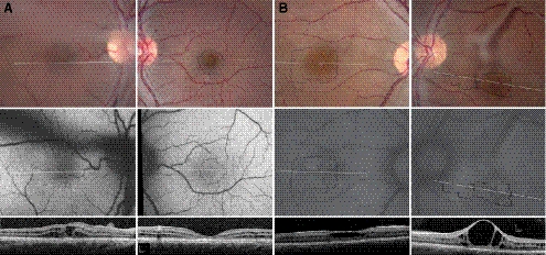
Figure 1.
Color (top), AF (middle), and SD-OCT images (bottom) of the right (left column) and left (right column) eyes of XLRS patient 1 (A) and patient 2 (B). Locations of the AOSLO images (Figures 2–4) are indicated by the thin, black outlines. SD-OCT locations are shown by the horizontal white lines. Vitreous veils created dark shadows visible in AF images of the right eye of patient 1 and the left eye of patient 2 and precluded acquisition of AOSLO images in the right eye of patient 1.

