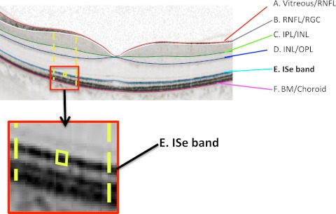
Figure 1.
An fdOCT horizontal scan through the fovea of a healthy control subject. The layers and bands manually segmented are indicated. Inset, the ISe band is shown without the segmentation line. BM, Bruch's membrane; INL, inner nuclear layer; IPL, inner plexiform layer; OPL, outer plexiform layer; RGC, retinal ganglion cell; RNFL, retinal nerve fiber layer.

