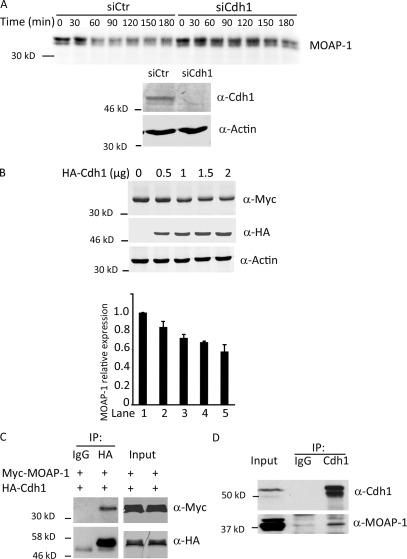
Figure 3.
Cdh1 regulates MOAP-1 stability. (A, top) HeLa cells were treated with control or Cdh1 siRNA, arrested in nocodazole, released for 4 h, and lysed, and lysates were supplemented with radiolabeled in vitro translated MOAP-1 protein. Samples were withdrawn into SDS loading buffer at the indicated times and resolved by SDS-PAGE and autoradiography. (bottom) Immunoblot of Cdh1 from the Cdh1 siRNA–treated (siCdh1) cells above. siCtr, siRNA control. (B) 293T cells were transfected with 0.5 µg Myc–MOAP-1 plasmid and increasing amounts of HA-Cdh1, as indicated. Cell lysates were collected and probed with anti-Myc, -HA, or -actin antibody. Relative Myc-MOAP1 levels (normalized to levels seen in the absence of exogenous Cdh1 in lane 1) are quantitated below based on three repetitions of the experiment. Error bars are SD. (C) 293T cells were transfected with 0.5 µg HA-Cdh1 and 1.5 µg Myc–MOAP-1. Cell lysates were prepared and immunoprecipitated (IP) by normal IgG or anti-HA antibody and probed with anti-HA or -Myc antibody. (D) Anti-Cdh1 or control IgG immunoprecipitates from 20 µM MG132-treated 293T cells were immunoblotted with anti–MOAP-1 or anti-Cdh1 antibody.

