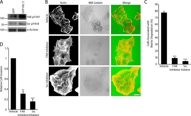
Figure 4.
FAK and Src activity are necessary for GFP–Hic-5–induced matrix degradation and invasion. (A) Western blot analysis shows increased levels of phosphorylated FAK and Src in GFP–Hic-5–expressing MCF10A cells compared with GFP cells. Molecular mass standards are indicated next to the gel blots in kilodaltons. (B) GFP–Hic-5 cells plated in the presence of FAK (PF573228) and Src (PP2) inhibitors fail to degrade matrix. Bar, 20 µm. (C) Quantitation of the percentage of cells associated with matrix degradation. (D) Matrigel invasion is significantly decreased in GFP–Hic-5–expressing cells treated with the FAK and Src inhibitors. Error bars represent the standard error of the mean. **, P < 0.005; ***, P < 0.0005.

