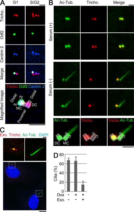
Figure 1.
Trichoplein disappears from the basal body in quiescent cells. (A) RPE1 cells were stained with antitrichoplein (Tricho.), anti-Odf2, and anti–Centrin 2. Bottom micrograph is shown with an illustration, which indicates the structure of a mother or daughter centriole (MC or DC) with the appendage (Ap) and orientations. (B) RPE1 cells were incubated in a medium with (+) or without (−) serum and then subjected to immunofluorescence with antibodies against acetylated tubulin (Ac-Tub.) and trichoplein. (C and D) Proliferating Tet-ON RPE1 cells expressing MBP-trichoplein-Flag (Tet-FL) were incubated in a new growing medium containing 30 ng/ml doxycycline (Dox) for 4 h and then cultured in a new serum-free medium containing 30 ng/ml Dox for an additional 44 h. (C) Cells were subjected to immunostaining with anti-MBP (Exo. tricho.) and anti–acetylated tubulin or immunoblotting (Fig. 7 E). (top) Magnified insets are shown. (D) The quantification of ciliation was shown in the graph. Exo. shows the centrioles with (+) or without (−) detectable exogenous trichoplein. We analyzed 100 cells per group and calculated the percentage of ciliated cells (n = 3). Data are means ± SD. Bars: (A–C, top) 1 µm; (C, bottom) 10 µm.

