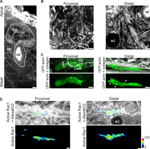
Figure 1.
Lobopodia-based 3D migration occurs in the mammalian dermis. (A) A 3D reconstruction of a mouse ear dermal explant labeled with Alexa Fluor 633 (grayscale). Stratum corneum (SC), basal keratinocytes (BK), papillary dermis (PD), and reticular dermis (RD) are indicated. Sebaceous gland (SG) and hair follicle (HF) are outlined in gray. (B) Examples of ECM structures proximal (left) and distal (right) to the basal surface of a dermal explant labeled with Alexa Fluor 633. Images are from the same confocal stack, 9 µm (left) and 30 µm (right) from the basal surface. AC, adipocyte. (C) 3D reconstructions of lobopodia-bearing HFFs migrating in proximal and distal collagen; GFP-actin is shown in green, and second harmonic imaging of collagen appears in grayscale. Arrowheads indicate lateral blebs. (D) Active Rac1 is not targeted to the leading edge of HFFs migrating in the mammalian dermis. Rac1 activity was imaged in HFFs migrating in proximal or distal ECMs; active Rac1, representing the Fc image, was pseudocolored according to the 16-color scale shown to the right of the figure, and the explant was labeled with Alexa Fluor 633 (grayscale). All cells are oriented with the leading edge toward the right of the figure. Bars, 5 µm.

