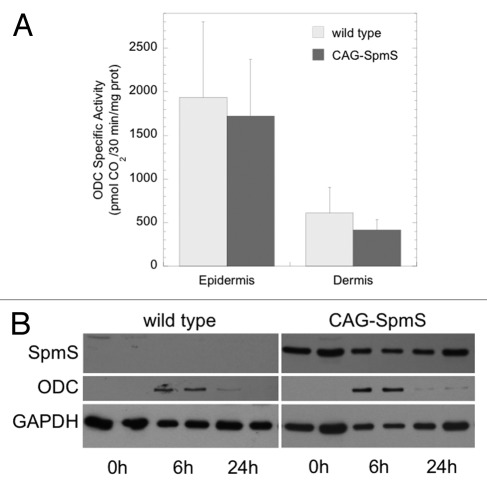
Figure 1. TPA-induced ODC activity and protein in wild type and CAG-SpmS mice. (A) ODC activity in wild type and CAG-SpmS mice treated with TPA (17 nmol). Mice were sacrificed 6 h after TPA application and assayed for epidermal and dermal ODC activity (mean ± S.D.; n = 4). (B) Epidermal extracts (50 µg) from wild type and CAG-SpmS mice were collected at the indicated time after TPA application (17 nmol) and duplicate samples were analyzed by western blotting for SpmS and ODC with GAPDH as a loading control.

An official website of the United States government
Here's how you know
Official websites use .gov
A
.gov website belongs to an official
government organization in the United States.
Secure .gov websites use HTTPS
A lock (
) or https:// means you've safely
connected to the .gov website. Share sensitive
information only on official, secure websites.
