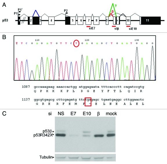
Figure 1. Characterization of p53 protein isoform expression in SK-N-AS cells. (A) Diagram of p53 gene showing coding exons (white boxes), non-coding exons (black boxes), the internal p53 promoter (Δ133 isoforms) and the β and γ splicings. The position of siRNA against Exon 7 (siE7), the β splicing of p53 (siβ) and exon 10 (siE10) are marked. (B) Sequence showing the single nucleotide nonsense mutation that results in the truncated p53R342X mutant present in SK-N-AS cells. (C) Western blot showing both forms of p53 detected in SK-N-AS cells. Cells are transfected with non-specific siRNA (siNS), siE7 (which targets both p53β and p53R342X), siE10 (which only targets p53R342X), siβ (which only targets p53β) and mock transfected, as indicated. Tubulin was used as a loading control.

An official website of the United States government
Here's how you know
Official websites use .gov
A
.gov website belongs to an official
government organization in the United States.
Secure .gov websites use HTTPS
A lock (
) or https:// means you've safely
connected to the .gov website. Share sensitive
information only on official, secure websites.
