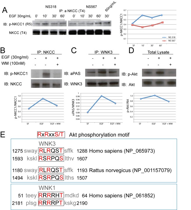
Figure 8. EGF promotes phosphorylation of NKCC1, and activation of the PI3K-Akt pathway is necessary for EGF-mediated WNK3 phosphorylation.
(A) Treatment of GB cell lines NS 318 and NS 567 with EGF (30 ng/ml) stimulates phosphorylation of NKCC1. Exposure of cells to EGF (30 ng/ml) for 10, 30, and 60 min show a time-dependent course of NKCC1 phosphorylation. Also, exposure of NS 567 to 60 ng/ml of EGF shows higher levels of phosphorylation than phosphorylation levels at the same time point at 30 ng/ml showing a dose-dependent effect. A line plot is presented with the quantification of the ratio of p-NKCC1/NKCC1. (B) Activation of PI3K is necessary for phosphorylation of NKCC1 after stimulation of HEK-293 cells with EGF. HEK-293 cells were serum starved overnight and were incubated with wortmannin (WM) for 30 min prior to stimulation with EGF for 30 min. Total cell lysate (150 µg) was immunoprecipitated with T4 antibody before immunoblotting with anti-phospho NKCC1 antibody (top panel) or T4 antibody (bottom panel). (C) Activation of PI3K is necessary for increased phosphorylation of WNK3 after stimulation with 30 ng/ml EGF. After overnight serum starvation, HEK-293 cells were incubated with WM for 30 min prior to stimulation with EGF for 30 min. Total cell lysate (150 µg) was immunoprecipitated with WNK3 antibody before immunoblotting with anti-phosphorylated Akt substrate (αPAS) antibody (top panel) or WNK3 (bottom panels). (D) Total cell lysate samples (25 µg) were also resolved by SDS-PAGE and blotted with phospho-Akt (threonine 473, top panel) and Akt (bottom panel) antibodies to show inhibition of Akt phosphorylation after PI3K inhibition. A line plot is presented with the quantification of the ratio of p-NKCC1/NKCC1, αPAS/WNK3, and p-Akt/Akt. (E) Akt phosphorylation motif, top panel. Middle panel shows the alignment of the sequences of rat and human WNK3 showing conservation of the Akt phosphorylation motif. Bottom panel shows sequence of WNK1, which is phosphorylated by Akt (bottom panel) [29]. Conserved residues are highlighted in red letters.

