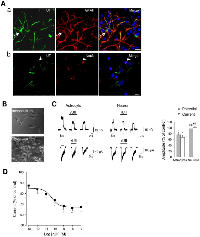
Figure 1. UII-induced depression of GABAAR in UT-expressing cerebellar astrocytes.
(Aa, Ab) Double immunofluorescence labeling of UT (green) and the specific astrocyte marker GFAP (red, Aa), or the mature neuron marker NeuN (red, Ab) in astrocyte-neuron co-culture from P7 rat cerebellum. Astrocytes, recognized by strong GFAP staining show UT immunoreactivity (arrows), whereas few weaker UT-stained cells express NeuN (arrowheads), and were likely attributed to mature granule cells (arrowheads, Ab). Nuclei (blue) were counterstained with DAPI. Scale bars, 50 µm. (B) Phase contrast photomicrograph of astrocytes in mono-culture, or astrocytes and neurons in co-culture at 3 days in vitro. (C) Membrane depolarizations and currents evoked by the GABAAR agonist isoguvacine (Iso, 10−4 M, 2 s for membrane potential and 5 s for chloride current) in astrocytes and cerebellar granule neurons before, during rUII (10−7 M, 40 s) application and after 2-min washout. Right, normalized amplitudes deduced by the mean Iso-evoked depolarization or current obtained before rUII application. (D) Concentration-response relationship of Iso-evoked currents from astrocytes yielding an EC50 value of 43.6±23.7 10−12 M. Data are mean ± SEM of 4 to 6 cells. *, P<0.05; ** P<0.01 compared with the corresponding control Iso-evoked current.

