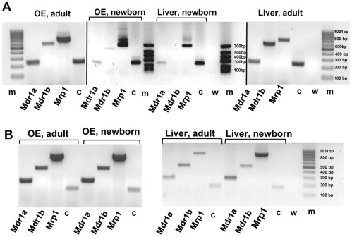
Figure 5. Distribution of mdr and mrp mRNAs in rat and mouse olfactory and liver tissue.
The presence of mdr1a, mdr1b and mrp1 mRNA analyzed by means of RT-PCR from samples of olfactory mucosa and liver tissue. Electrophoresis of PCR products A: from newborn and adult rat, B: from newborn and adult mouse. The cDNAs were amplified with rat- and mouse-specific primers for the Mdr1a, Mdr1b and Mrp1 genes. All tissues were positive for the tested cDNAs (note that staining in A, lane 12 for mdr1b is weak, but visible on the gel). c = cyclophilin A used as positive control, w = negative control containing water in place of cDNA, m = DNA size markers. All gels were run together except for the newborn rat, which was run on a separate gel. Results from a single experiment.

