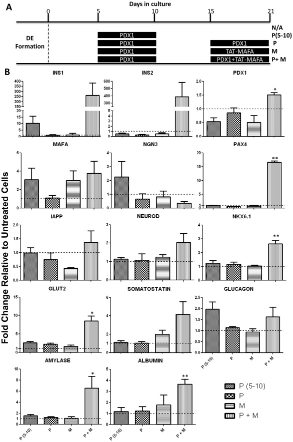
Figure 4. Pdx1 and TAT-MafA promote pancreatic differentiation of DE-enriched mouse ES cells.
(A) Diagram of the different differentiation culture conditions. After definitive endoderm (DE) formation mES cells were plated as a monolayer and treated for 21 days with the indicated combinations of Pdx1 (P) and TAT-MafA (M). (B) On day 21 cells were harvested and total RNA was extracted for analysis of the expression of pancreatic genes by RT-qPCR. Data represent the average of triplicate experiments and are expressed as mean ± SEM. Data are expressed as gene expression relative to GAPDH and normalised against untreated (N/A) cells, where p<0.05 (*), p<0.01 (**) or p<0.001 (***).

