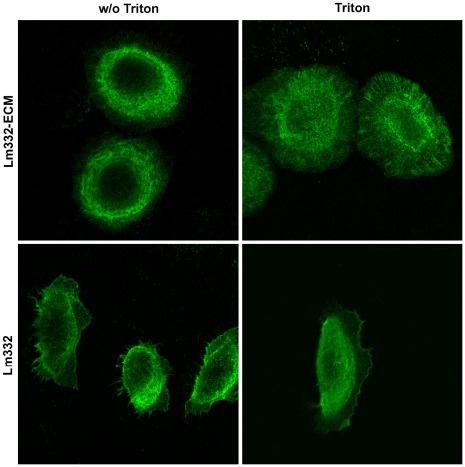
Figure 10. Localization of integrin ß4 in NHK cells on purified Lm332 and Lm332-ECM.
Glass-bottom dishes (Asahi Techno Glass, Tokyo, Japan) were previously coated with 2.0 µg/ml Lm332 or deposited with Lm332-ECM. NHK cells were inoculated and incubated in growth media. After incubation for 5 h, the cells were washed with PBS and then fixed with 4% (w/v) paraformaldehyde in PBS for 10 min and then treated without (w/o Triton) or with (Triton) 0.5% (v/v) Triton X-100. The fixed cells were stained with an integrin ß4 antibody and an Alexa Fluor 488-labeled secondary antibody. Other experimental conditions are described in “ Materials and Methods”.

