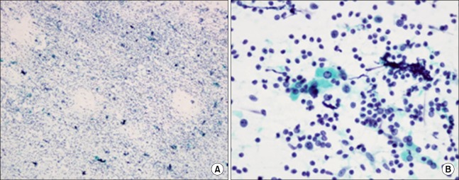
FIG. 3.
The cytopathology of the thyroid gland. (A) The cytopathology of the thyroid gland revealed reactive and polymorphous lymphoid cell infiltrations (Papanicolaou's stain, ×100). (B) Occasional epithelial cells with Hürthle cell changes were mixed with lymphocytes, centroblasts, plasma cells, and macrophages (Papanicolaou's stain, ×200).

