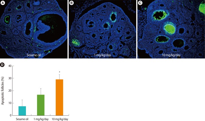
Figure 2.
Detection of apoptotic cells in the ovary after tributyltin (TBT) administration (A-C). Paraffin sections of week-old rat ovaries were stained by using a terminal deoxynucleotidyl transferase dUTP nick end labeling (TUNEL) assay kit and counter-stained with 4',6-diamidino-2-phenylindole after TBT exposure for 7 days. Apoptotic cells stained by TUNEL show as green. ×200. (D) The percent of apoptotic follicles counted visibly increased in the ovary after TBT exposure.

