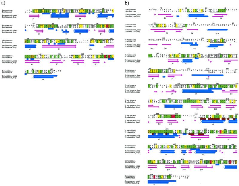
Figure 1.
Sequence alignments of AS subunits from S. marcescens and S. solfataricus calculated from aligned structures of the two molecules. Structure alignment was performed with o (24) and stamp (36) and the figure produced by alscript (37). Secondary structures were assigned by dssp (38). Numbering is based on the S. marcescens sequence. Conserved hydrophobic residues are shaded yellow, conserved nonhydrophobic residues are shaded green, conserved polar residues are in bold font, conserved small residues in small font, and conserved structural regions are shown boxed. Those residues underlined in a shaded blue box are solvent inaccessible in the heterodimer TrpE:TrpG interface, whereas those in brown are solvent excluded in the heterotetramer interface. β-Sheet regions are shown as light purple arrows, whereas α-helices are in blue. (a) Alignment of TrpGs: residues of the active site are shaded red, whereas those in close contact with the glutamyl residue are shaded gold. (b) Alignment of TrpE: residues in close contact to anthranilate shaded red, whereas those in close contact with pyruvate are in light blue. Residues shaded gold are in close contact with tryptophan.

