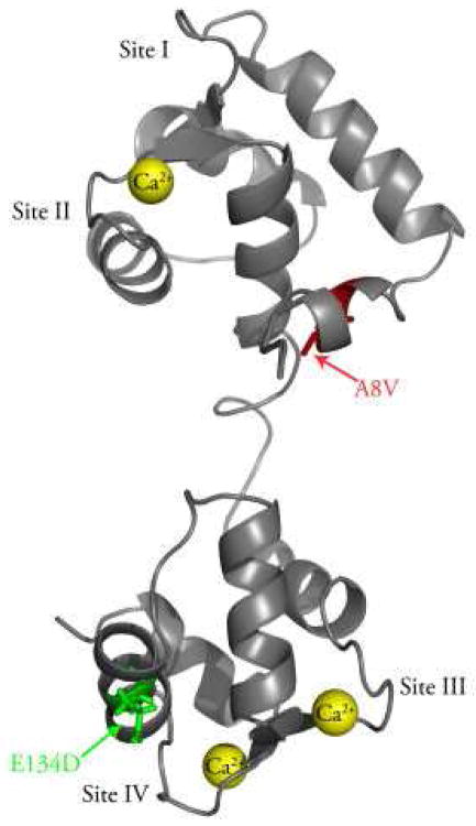
Figure 1. Location of the A8V and E134D mutations in the N- and C-domains of cTnC.
The figure shows a ribbon representation of cTnC in the Ca2+ bound state (Protein Data Bank entry 1AJ4 (37)). The A8V mutation (shown in red) is located in the N-helix of the N-domain, while the E134D mutation (shown in green) is located between Ca2+ binding sites III and IV. This figure was generated using PyMOL (www.pymol.org).

