
Figure 1.
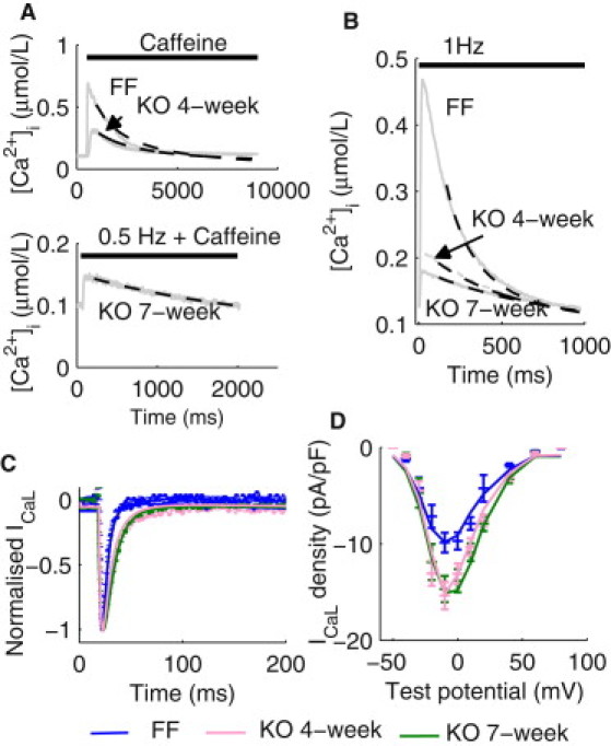
Parameterization of Ca2+ handling mechanisms. (A) (Top panel) Representative caffeine-induced [Ca2+]i transients in isolated ventricular myocytes from the FF and KO four-week mice. Simulated decay of the [Ca2+]i transients (dashed lines) using the respective final fitted parameter values for NCX and PMCA are superimposed. (Bottom panel) A representative [Ca2+]i transient paced at 0.5 Hz in the presence of caffeine in the KO seven-week cardiomyocytes. Simulated decay of the [Ca2+]i transient (dashed line) using the final fitted parameter values for NCX and PMCA are superimposed. (B) Representative [Ca2+]i transients at 1 Hz measured in the FF, KO four-week, and KO seven-week cardiomyocytes. Simulated decay of the [Ca2+]i transients using the respective final set of NCX, PMCA, and SERCA parameters are superimposed. (C) Representative normalized ICaL time courses under voltage-clamp from −50 mV holding potential to −10 mV test potential in the FF, KO four-week, and KO seven-week cardiomyocytes (dotted lines). Simulated ICaL timecourses under the voltage-clamp protocol are superimposed (solid lines). (D) Current-voltage relationship of ICaL in the FF, KO four-week, and KO seven-week cardiomyocytes. Simulated I/V relationships are superimposed.