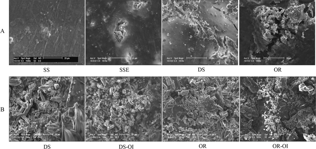
Fig. 3.
Scanning Electron Micrograph of the mineral nodule formation of ASCs grown on stainless steel (SS), etched stainless steel (SSE), disordered (DS) and ordered (OR) FA surfaces after osteogenic induction (OI) for 4 weeks. The mineral deposition was beneath the densely formed cell-matrix layer (A). After removal of the cells, more densely deposited amorphous mineral nodules were observed on the ordered FA surfaces with and without the OI (B).

