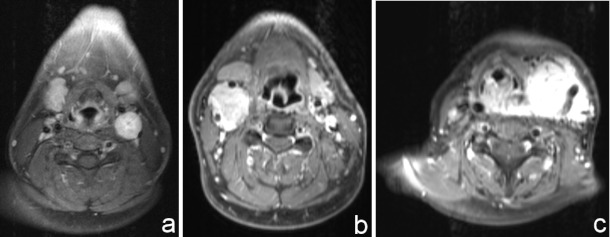
Figure 1. Figure 1a: CBT (class I): The axial T1-weighted MRI with contrast medium shows a CBT in typical location. Splaying of the left carotid bifurcation without any surrounding of the carotid vessels.
Figure 1b: CBT (class II): Axial T1-weighted MRI with contrast showing a right-sided CBT partially surrounding the internal and external carotid artery.
Figure 1c: CBT (class III): A large left-sided CBT intimately surrounding the carotid vessels (axial T1 weighted MRI with contrast).

