
Fig. 6.
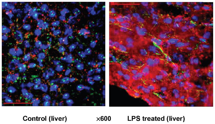
iNOS recorded by immunofluorescence staining in the liver in control pigs (without LPS) and treated with LPS at 200 μg kg−1 over 60 min
Blue indicates 4′,6-diamidino-2-phenylindole (nuclei); green, smooth muscle actin (vasculature); red, iNOS.