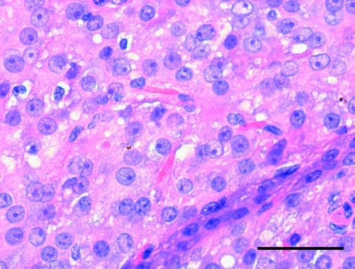
Figure 1.
Standard H&E-stained section (5 μm) of testicular Leydig cell tumour. The intra-testicular tumour (largest diameter of 1.8 cm) was sharply delimited from the testicular parenchyma. The tumour cells had abundant, deeply acidophilic, finely vacuolised cytoplasm with focally deposited brownish yellow lipochrome pigment and intracytoplasmic Reinke's crystalloids. Scale bar=50 μm.

