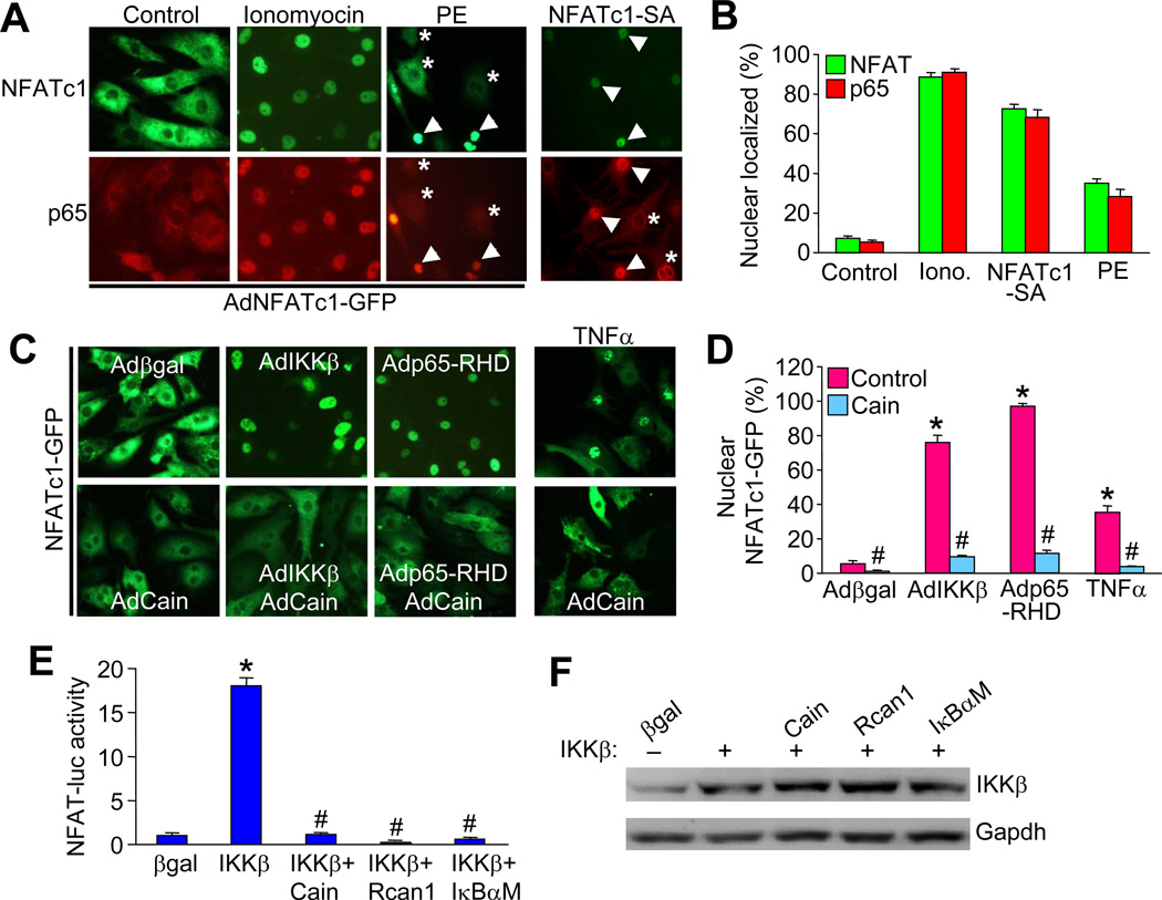
Figure 3. Co-localization of NFAT and NFκB in cardiomyocytes.
A, Immunofluorescent images from cultured neonatal cardiomyocytes infected with AdNFATc1-GFP (green) and stimulated with 1 µM ionomycin, 50 µM phenylephrine, or vehicle control, or infected with an NFATc1-SA-GFP adenovirus. Cells were also immune-stained for endogenous p65 (red). Arrowheads indicate nuclear localization while asterisks show cytoplasmic localization. B, Quantification of nuclear localized NFATc1 and p65 in cells treated as indicated in A. C, Immunofluorescent images from cultured neonatal cardiomyocytes infected with AdNFATc1-GFP along with the other indicated adenoviruses or stimulated with 20 ng/ml TNFα. D, Quantification of nuclear localized NFATc1 in cells treated as indicated in C. E, NFAT-luciferase activity in neonatal cardiomyocytes infected with the AdNFAT-luciferase reporter and the other indicated adenoviruses. *P < 0.05 versus β-gal; #P < 0.05 versus IKKβ only. Results were summated from 3 independent experiments. F, Control western blot to show equal levels of IKKβ overexpression for the experiment shown in E.

