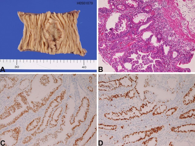
Figure 1.
Adenocarcinoma of the small intestine. A: Macroscopic picture in the ileum. An ulcerated tumor is present. B: Histology of well differentiated adenocarcinoma of the duodenum. HE, x200 C: p53 expression in adenocarcinoma of the duodenum. D: Ki-67 expression in adenocarcinoma of the duodenum. The labeling is 90%, x100.

