Figure 1.

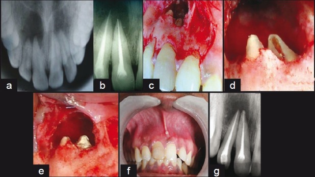
(a) Previous occlusal radiograph, (b) preoperative IOPA radiograph, (c) exposed lesion, (d) exposed apical transportation, (e) defect repaired by MTA, (f) third month recall, (g) recall radiograph

(a) Previous occlusal radiograph, (b) preoperative IOPA radiograph, (c) exposed lesion, (d) exposed apical transportation, (e) defect repaired by MTA, (f) third month recall, (g) recall radiograph