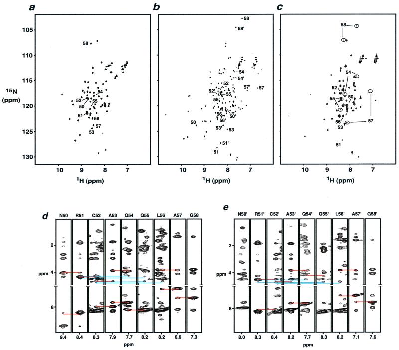
Figure 4.
1H-15N heteronuclear single quantum correlation (HSQC) spectra of dimer HP62-V52C (a) in the free form, (b) in the complex with wild-type DNA operator, and (c) in the complex with SymR(+1) operator. Peaks corresponding to the hinge region residues are labeled. (b) Normal numbers indicate residues of the left site, whereas primed numbers indicate residues of the right site. (c) Peaks corresponding to the different folding states of hinge region residues are indicated in circles. Strip plots of residues 50–58 from (1H-15N) NOESY-HSQC spectra of HP62-V52C–wild-type DNA complex: (d) left site, (e) right site. Hα–HN and HN–HN sequential contacts are indicated with red lines; Hα–HN(i, i + 3) and Hα–HN(i, i + 4) contacts are indicated with blue lines.

