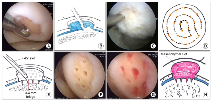
Fig. 1.
Surgical procedure of microfracture. (A) Unstable cartilage flap and calcified cartilage bed is debrided with open curette. (B) It is important to debride the calcified cartilage layer and make a well-contained pocket surrounded healthy cartilage (well-shouldered). (C) Subchondral bone is punctured with an awl. (D) Microfracture is circumferentially performed from periphery to center. (E) The penetration of subchondral bone is 3 to 4 mm deep and apart. (F) Arthroscopic photograph showing the final step of microfracture. (G) Mesenchymal blood egress from bone marrow through subchondral holes. (H) It is important for tissue regeneration to keep the mesenchymal clot in the defect.

